


HistoLab Image Analysis Software for Fully Automatic Feature Size, Shape and Colour Analysis
Couldn't load pickup availability


HistoLab was originally developed to take several types of challenging measurements of various types of histological tissue. As a result it includes a diverse range of features for processing images and numerous interactive and powerful measurement tools.
We have taken particular care with regards to simplicity and ease of use and we wanted the documents produced to be usable straight away: incorporating a printout of the images and the results, storage in files that can be manipulated with other applications and statistics on all of the analysed structures.
HistoLab is designed to provide the user with immediate, easy access to a wide variety of measurements on diverse types of histological preparations.
HistoLab’s sophisticated image processing functions and many powerful, interactive measurement facilities make this image analysis system an essential tool for the modern histologist.
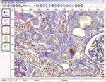
Histolab™ allows you to quickly and accurately analyze all tissues in a histological section and any cells and subcellular structures visible under a microscope.
The results of the statistics and measurements are shown in the table (counting, length, width, diameter, perimeter, surface, intensity) are expressed in real units in complete tables
When fitted with an encoder stage, Histolab™ is not limited to the microscope's current field of view : all measurements can be located over the entire section.
Dedicated image analysis software has been developed for the analysis of micronuclei, the estimation of the cellular cycle by autoradiography, and the quantization of the activity of cells by a mitogen
HistoLab is normally supplied wth an integrated camera, Nazca camera interface software and often with motorised stage and focus systems for your microscope.
Shipping is by courier and takes typically 2-5 days but this can alter.
Please phone us on +44 (0)1284 789697 or email us for current delivery times.
Request a quotation for bulk orders by emailing sales@gtvision.co.uk
If you have any questions about this model, complete the form below or email us at sales@gtvision.co.uk
- Because we supply microscopes from a wide variety of brands and manufacturers, we can offer expert advice on which microscope model is best suited for your application and budget without bias towards a particular brand.
- We provide tailor-built systems to match your specific requirements and budget.
- Expert support pre and post purchase.- For a full system solution, we also supply microscope cameras and software (again from a wide choice of brands).
- We can retro-fit cameras and illumination systems, including modern LED fluorescence illuminators, to most models and ages of existing microscopes. We also have in-house engineering capabilities for particularly unique requirements.