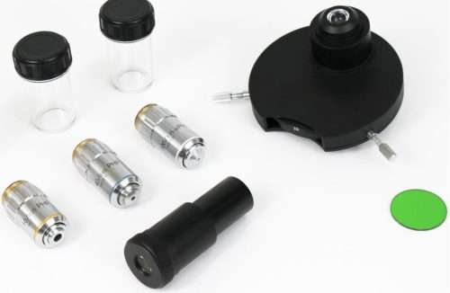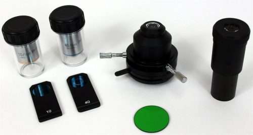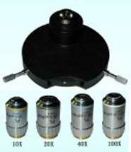
See here our range of phase kits for GXM biological upright microscopes, all of our inverted biological microscopes have phase contrast outfits included as standard. If you need a phase acessory for another microscope model (eg Leica,Olympus, Nikon etc) please let us know, we will be pleased to help.
Phase contrast microscopy is an optical microscopy technique that converts phase shifts in light passing through a transparent specimen to brightness changes in the image. Phase shifts themselves are invisible, but become visible when shown as brightness variations.
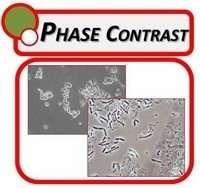
 Figure 1:
 The same cells imaged with traditional bright field microscopy (left) and with phase contrast microscopy (right).
When light waves travels through a medium other than vacuum, interaction with the medium causes the wave amplitude and phase to change in a manner dependent on properties of the medium. Changes in amplitude (brightness) arise from the scattering and absorption of light, which is often wavelength dependent and may give rise to colors. Photographic equipment and the human eye are only sensitive to amplitude variations. Without special arrangements, phase changes are therefore invisible. Yet, phase changes often carry important information.
Phase contrast microscopy is particularly important in biology. It reveals many cellular structures that are not visible with a simpler bright field microscope, as exemplified in Figure 1. These structures were made visible to earlier microscopists by staining, but this required additional preparation and killed the cells. The phase contrast microscope made it possible for biologists to study living cells and how they proliferate through cell division.[1] After its invention in the early 1930s, phase contrast microscopy proved to be such an advancement in microscopy, that its inventor Frits Zernike was awarded the Nobel prize (physics) in 1953.
Working principle
The basic principle to make phase changes visible in phase contrast microscopy is to separate the illuminating background light from the specimen scattered light, which make up the foreground details, and to manipulate these differently.
The ring shaped illuminating light (green) that passes the condenser annulus is focused on the specimen by the condenser. Some of the illuminating light is scattered by the specimen (yellow). The remaining light is unaffected by the specimen and forms the background light (red). When observing unstained biological specimen, the scattered light is weak and typically phase shifted by -90° — relative to the background light. This leads to that the foreground (blue vector) and the background (red vector) nearly have the same intensity, resulting in a low image contrast (a).
In a phase contrast microscope, the image contrast is improved in two steps. The background light is phase shifted -90° by passing it through aphase shift ring. This eliminates the phase difference between the background and the scattered light, leading to an increased intensity difference between foreground and background (b). To further increase contrast, the background is dimmed by a gray filter ring (c). Some of the scattered light will be phase shifted and dimmed by the rings. However, the background light is affected to a much greater extent, which creates the phase contrast effect.