Free Delivery On Orders Over £200 to UK Mainland

01284789697

Our Top 13 Microscopy Influencers To Follow

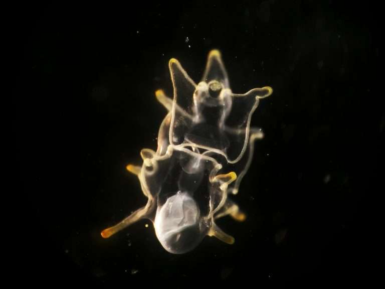
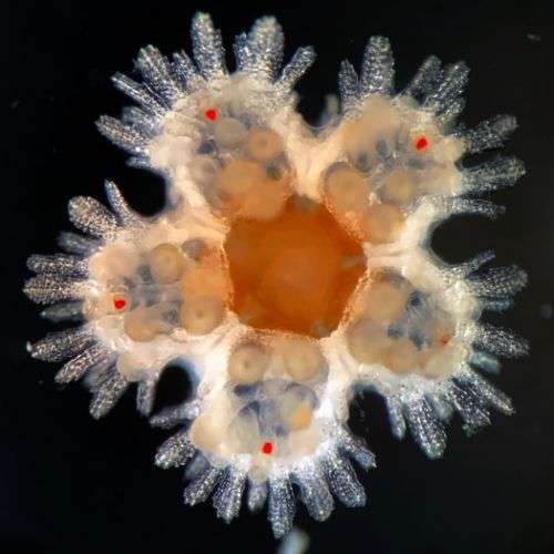
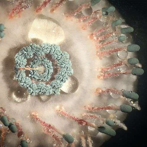
Starfish larva
Starfish larva, captured by Igor Adameyko

With rapid advances in microscopy imaging and mobile phone camera technology over recent years, the micro world has been making its way out to the bigger world of social media and YouTube. The response has been incredible, with several microscopy-related accounts reaching over 250,000 followers and some even nearing the million mark!

Instagram, Twitter and TikTok feeds plus YouTube channels are being filled with visually stunning and educational videos, covering a large range of topics from soil microbiology to veterinary cytology and everything in between.

We’ve compiled a list of our top 13 microscopy influencers to share with you, so grab a cup of tea and prepare to be glued to your screens in awe as you explore the fascinating and beautiful microscopic world through social media…

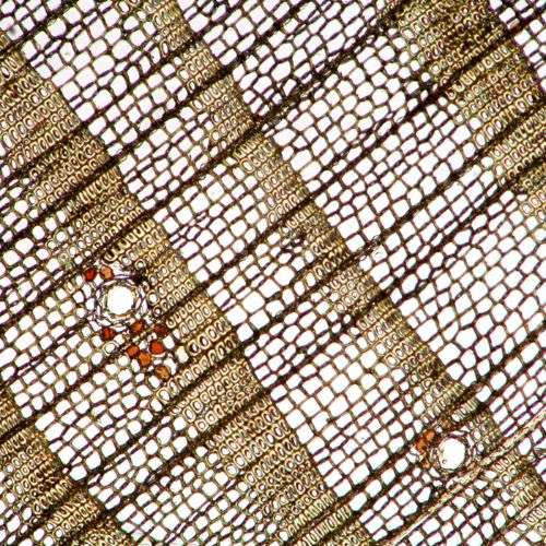
microbehunter spruce wood under microscope
Spruce wood section
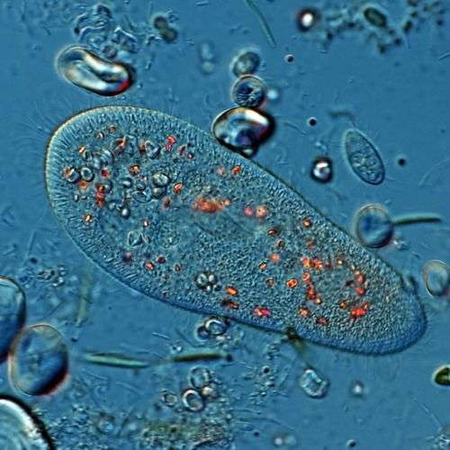
Paramecium under the microscope by Microbehunter
Paramecium
microbehunter microscopy
The Microbehunter Microscopy website

OLIVER | @MICROBEHUNTER

Subject matter: the environment and nature

Microscope setup: I have several microscopes from different brands. I generally take pictures using a DSLR camera connected to the phototube but also use a USB microscope camera. I also use the DSLR for live-viewing on a computer monitor and for live-streaming. This camera can also be connected directly to a monitor without a computer. 

“I am Oliver and am known in social media under the name “Microbehunter”. My mission is to encourage people to observe their environment and nature with a microscope. Of course, this includes microorganisms but it is not limited to that. Especially plants can be quite pretty under the microscope. In short, I want to make the hobby of Amateur Microscopy more popular.

I think that a solid (also theoretical) formal science education has a lot of merit in understanding how our world works, but often there are simply not enough resources (time!) available during education to explore things hands-on. With my YouTube videos I want to motivate people to pick up a microscope for themselves and to start exploring their environment. I have heard that teachers also show my videos in class to spice up their lessons.

Now a few words about my background. I have a Master’s degree in Microbiology and a teacher’s education license for Biology and Information Technology. For this reason, the microscopic world has been part of my life for a long time. As a teacher, I am teaching my students the theoretical foundations of Biology and microscopic observation. With my YouTube Channels, I am reaching out to also an older audience, hoping to get them excited about the microscopic world as well.

While I have worked with microscopes on and off since university, I only became really active in 1998, when I bought my first (good) microscope. At that time affordable microscope cameras were not available. I experimented with several DIY solutions, including connecting a surveillance camera and webcams.

In 2011 I started to publish a free monthly microscopy magazine (still available for download on my website). After a few years, I found out that by posting YouTube videos I am able to reach a far larger audience and I have been publishing YouTube videos since 2018.

I have a website with information, and several YouTube channels about microscopy, two of which are quite active. The larger channel of the two is dedicated to observing nature under the microscope. It’s focussed mainly on the specimens. The other channel is a bit more technical, featuring microscope reviews, advice, questions & answers, and a weekly livestream.

For those of you, who consider to start microscopy as a hobby, I have the following advice: Do not spend too much at the beginning, but buy new microscopes as you grow your experience. This way it will be much easier for you to make a choice that is best for you. And do not forget that one central point of hobby microscopy is to observe nature. The microscope is only a tool to achieve this goal.

Happy microbe hunting! Oliver”

Find Oliver on:

Website: www.microbehunter.com

YouTube channel on observing with a microscope: @microbehunter 

YouTube channel with reviews, technical info & livestream: @microbehuntermicroscopy

QUINTEN GELDHOF | @MICROHOBBYIST

Subject matter: the natural world

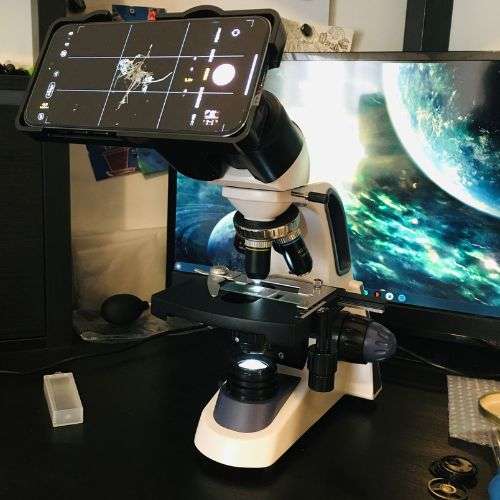
Microscope setup: Swift 380B microscope with iPhone 14 Pro attached via an iLabCam adapter

“My name is Quinten Geldhof and I’m a 23 year old Mechanical engineer from Massachusetts USA with a love for the natural world and microscopy. I’m relatively new to the microscopy scene and only got my first microscope in June of 2022. I was inspired to get one through the fantastic microscope streams by Faezaria on Twitch and the videos by AVNJ and Journey to the Microcosmos on YouTube, all of which blew me away with the complexity and beauty of the microscopic world.

I use a Swift 380B microscope combined with an adapter from iLabcam which which was a budget friendly choice to get into the hobby and has been great way to learn the ropes and still capture beautiful imagery.

I like to look at whatever I think might be interesting under the microscope which is a seemingly infinite number of things. This includes samples from fresh and salt water environments, various produce items, plants, crystals, and even my own blood. I love researching and sharing about the samples I post and there is always so much to learn about and I’m grateful to all those who share in the excitement with me.”

Find Quinten on:

Instagram: @microhobbyist 

Our Top 13 Microscopy Influencers To Follow
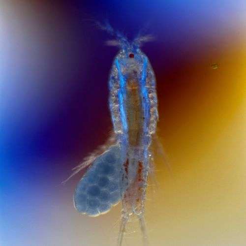
Copepod captured using polarised light
Our Top 13 Microscopy Influencers To Follow
Quinten's Swift 380B Microscope with iLabCam Adapter
Our Top 13 Microscopy Influencers To Follow
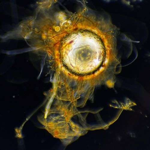
Air bubble trapped in algae
Our Top 13 Microscopy Influencers To Follow
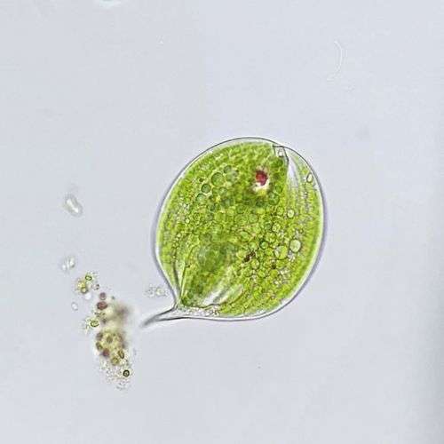
Phacus of the phylum Euglenozoa
Darkfield microscopy
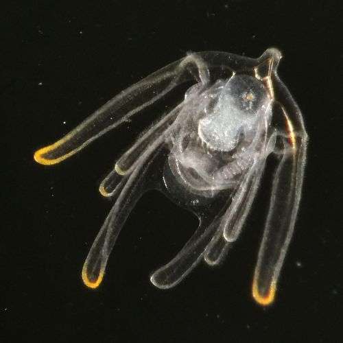
Post-metamorphic starfish using darkfield microscopy
Brittle Star larva using darkfield microscopy
Brittle Star lava using darkfield microscopy

IGOR ADAMEYKO | @THE_STORY_OF_A_BIOLOGIST

Subject matter: marine biology

Microscope setup: Zeiss Standard 14 Compound Microscope and Olympus SZ30 Stereo Microscope with iPhone attached via a Celestron adapter

Igor Adameyko, a professor of biology, runs YouTube (@igor_adameyko), Instagram (@the_story_a_biologist) and TikTok (@igoradameyko) accounts focusing on marine biology, with particular emphasis on the diversity of planktonic life forms. Igor uses a variety of microscopy techniques, including darkfield microscopy and confocal microscopy, to showcase the exquisite features of small marine animals that belong to different groups on the tree of life. These groups include mollusk larvae, tiny jellyfish, copepods, polychaetes, chaetognaths, amphipods, and many more; sometimes even parasites found on planktonic animals. 

As a keen diver and explorer with a sharp eye for detail, Igor developed a fascination for marine biology and the life cycles of marine animals around a decade ago. He began collecting old microscopes to create mobile setups for darkfield videography, which he can take with him in a backpack to various locations. These setups, which are based on Zeiss Standard 14 and Olympus SZ30, are still in use today. Igor attaches his iPhone to the microscope via a Celestron adapter and records the animals on rocks, in hotel rooms, or at biological stations. 

The sea is truly boundless, and exploring the marine world on a microscale is crucial for conservation efforts and comprehending the circle of life in the ocean.

Professional biologists and the general public alike will find Igor’s videography of minuscule creatures, barely visible to the naked eye, fascinating and accompanied by in-depth descriptions.

Find Igor on:

Instagram: @the_story_of_a_biologist

YouTube: @igor_adameyko

TikTok: @igoradameyko

MARTIN KAAE KRISTIANSEN | @MY.MICROSCOPIC.WORLD

Subject matter: mostly microscopic animals and single celled organisms but also enjoys looking at other things under the microscope

Microscope setup: Motic BA310E microscope with a Nikon z50 camera

“My name is Martin Kaae Kristiansen and I am from Denmark. I have a master’s degree in biomedicine. During my education I did some projects using microscopes and fell in love with the microscopic world. So I bought my own microscope and started looking at everything I could get my hands on. I was amazed at the incredible beautiful and alien like world only visible through a microscope. So I wanted to share it with as many people as possible. When I take a sample I use a lot of time researching the organisms I encounter so I can give a short, informative, and hopefully interesting, with my posts here on Instagram. Most of my posts are microscopic animals and single celled organisms I find I nature. But from time to time I include some human cells, crystals, and other stuff as well. Everything under a microscope of course. I want to make the invisible world visible for as many people as possible.”

Find Martin on:

Instagram: @my.microscopic.world

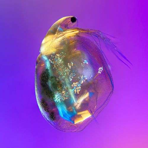
Simocephalus at 4x magnification
Simocephalus taken using the 4x objective lens
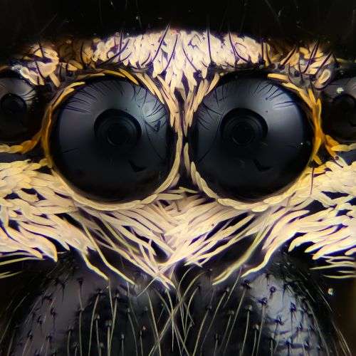
Jumping spider taken using the 10x objective lens
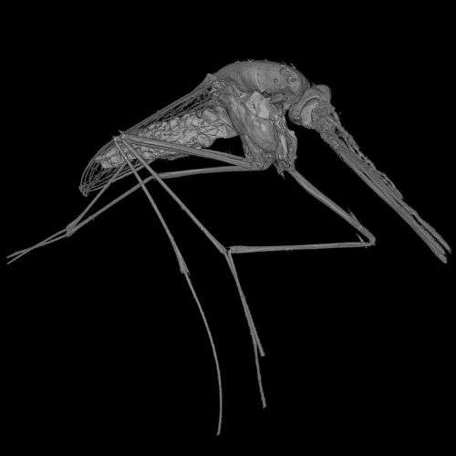
CT image of a mosquito
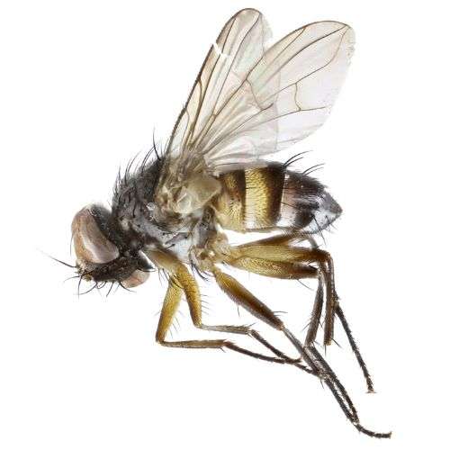
Pseudoredtenbacheria fly
New material caught in Dominia - Pseudoredtenbacheria
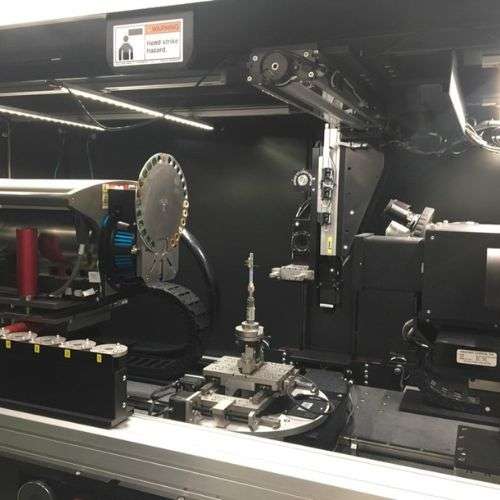
The Micro CT Scanner at the Natural History Museum
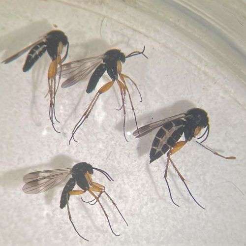
Fungus gnats
Fungus gnats - Greenomyia mongolica

DR. ERICA MCALISTER | @FLYGIRLNHM

Subject matter: entomology; specifically flies

Microscope setup: Leica MZ6 stereomicroscope with a Schott KL1500 compact illuminator 

“My twitter account is mostly flies. Flies from the collection at the Natural History Museum, flies seen or sampled in the field, and stories about flies that have piqued my interest.

I spend a lot of time studying and imaging flies so many of the images are macro or from imaging microscopes. I am obsessed about these ignored or maligned creatures and love to show off their form and variety.”

Find Erica on:

Twitter: @flygirlNHM

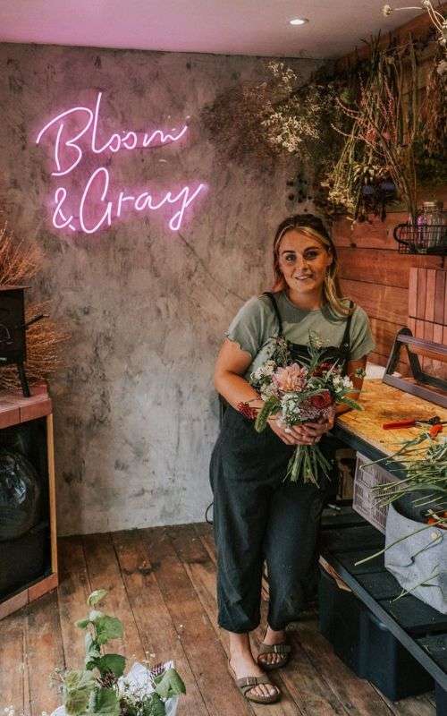
SARAH GRAY | @BLOOMANDGRAY

Subject matter: flowers, soil health and regenerative farming

Microscope setup: GX Microscopes L1500 compound microscope with GXCAM HiChrome-HD Camera 

“My fascination with soil biology came gradually, I started hearing things about soil health and regenerative farming in podcasts I was listening to and it really started to line up with how I wanted to manage my flower farm. I was always a little confused by synthetic fertilisers, herbicides and pesticides and stayed away from them out of caution. When I heard from other farmers who were working with nature to enhance their farms I was straight on board with them! It made so much more sense. Since then I have read lots of books, podcasts, YouTube videos and attended courses to learn as much as I can. I eventually took the plunge and decided to get a microscope to see what was going on inside my soil. It was so exciting to actually see much heard about members of the soil food web right in front of my eyes!

My Instagram and YouTube channel features various content relating to flower growing in the UK. I cover ‘how-to’s’, day to day life on the farm, lots of soil health and regenerative farming related stuff and some floristry content. I love sharing the ups and the downs and my learning experiences. I don’t want anyone to be under the illusion that flower farming is easy!”

Find Sarah on:

YouTube: @Bloomgrayflowerfarm

Instagram: @bloomandgray

Website: www.bloomandgray.co.uk

Regenerative farming with Bloom & Gray
Sarah Gray of Bloom & Gray
bloom and gray flowers
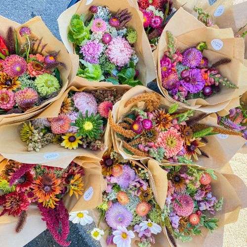
Flowers from Bloom & Gray
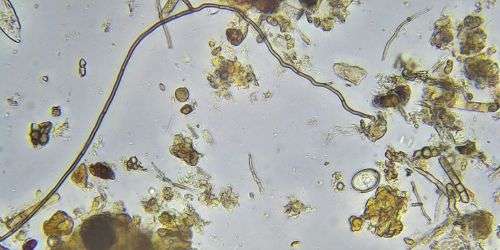
Fungal hyphae testate amoeba
Fungal Hyphae Testate Amoeba
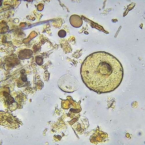
Arcella Amoeba
Arcella Amoeba

EDDIE BAILEY | @RHIZOPHYLLIA

Subject matter: soil food web microorganisms

Microscope setups: 

“I have been an organic allotmenteer for 30 years but 12 years ago I noticed my soil structure breaking down and plants suffering, not least in poor germination rates. I searched on-line and found Dr Elaine Ingham and the Soil Food Web. It dawned on me that my annual rotavating had been disrupting nature’s attempts to help and educate me! I stopped digging and ever since have been researching the microorganisms; today they do most of the work for me and I have healthier, more nutrient-dense crops.

In my Instagram and Twitter feeds we focus on the soil food web microorganisms (who they are and what substances and services they have evolved in symbioses with plants) in order to improve the outcomes of gardeners and growers of any scale in a sustainable and truly natural way. This includes posts on ground conditions, signs of a fully or poorly functioning soil food web, and ways to improve or rebuild your soil food web with composts and teas.”

Find Eddie on:

Instagram: @rhizophyllia

Twitter: @rhizophyllia

Website: www.rhizophyllia.co.uk

YouTube: @rhizophyllia5950

PROFESSOR ADOLFO SANCHEZ-BLANCO | @DR.BIO4EVER

Subject matter: biological samples under the microscope

Microscope setup: Olympus CX31 microscope and Leica Zoom 2000 stereo microscope

“I am a Spanish biologist established in the United States. I started my career as an Ecologist but then I became a graduate student in Biomedical Science. For about a decade after obtaining my Ph. D., and before I started my college teaching career, I worked as a full-time scientist. As part of my scientist career, I worked for 6 years at Stanford University investigating the molecular genetics of aging in the nematode worm C. elegans. Since this model organism is microscopic, I had to constantly use different types of microscopes to perform my research. This got me very excited into the microscopy world.

I am now a Biology professor at Capital Community College in Hartford, Connecticut.

When I started my teaching career and noticed that a number of topics that we would cover in class involved performing microscopy experiments, I decided to start posting Instagram and TikTok videos that would summarize these and other biology experiments in less than 1 minute. My intention was to show my Biology students that Biology can indeed be interesting and easy to understand. In other words, my main goal was to use the power of social media to excite my students to learn the material that we cover in class.

My Instagram account features microscopy and biology stories as well as all-about nature videos. In my videos, I try to make microscopy and biology relatable and showcase the beauty of understanding biology. For example, a number of my videos involve sampling remote ponds or streams of water in the forest and showing the amazing microscopic creatures that can be found there.

I recently opened a YouTube account with the intention of doing more in-depth microscopy and biology videos. I also, in collaboration with one of my professor colleagues at my college, published a biology lab manual that integrates QR codes to my Biology Instagram account to further illustrate students how to do the experiments that students need to do in the labs.”

Find Adolfo on:

TikTok: @dr.bioforever

Instagram: @dr.bio4ever

YouTube: @dr.bioforever2913

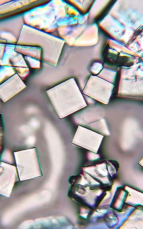
Ocean salt crystals under the microscope
Ocean salt crystals
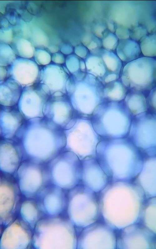
Broccoli under the microscope
Broccoli
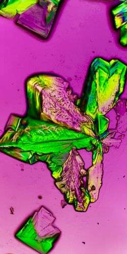
Absinthe under the microscope
Absinthe crystals
Our Top 13 Microscopy Influencers To Follow
Surface of a bubble
Our Top 13 Microscopy Influencers To Follow
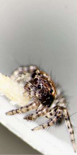
Baby jumping spider
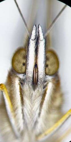
Butterfly face under the microscope
Butterfly face

WALT | @ONEMINMICRO

Subject matter: anything and everything viewed under the microscope!

Microscope setups: 

  • Nikon Eclipse E200 for biological samples
  • I have an unbranded stereoscope for some of the larger samples


“For as long as I can remember I’ve always loved microscopes and I’ve always been fascinated with life on a smaller scale. I’ve always marvelled at the thought of bugs and mini beasts, especially how they live, think and function! I am always wondering how small life gets before it becomes viewed as just a chemical reaction. It was only really in 2021 when I decided to publish my microscope findings onto social media as I enjoy sharing my findings with the world. Since then, I’ve found my voice as a microscope content creator/influencer and have amassed over a 2 million follower count across popular social media platforms TikTok (1.6M), Instagram (423k) and YouTube (546k).

If one visits my accounts, they should to expect to find anything and everything viewed under the microscope – I love to push the boundaries of conventional microscopy and have shown in the past some weird, wacky and wonderful things including, but not limited to: chemical explosions up close, jumping spiders, butterflies, lizards, frogs, various alcoholic beverages, meteorites, fossils as well as more traditional samples such as blood, microbes, and creek, puddle, ocean water samples. It’s anyone’s guess what I might post next!”

Find Oneminmicro on:

Instagram: @oneminmicro

TikTok: @oneminmicro

YouTube: @oneminmicro

BEN ALVERSON | @VETERINARY_PARASITOLOGY_

Subject matter: microscope photos from a vet diagnostic laboratory

Microscope setup: Olympus CH-2 compound microscope with standard 4x,10x, 40x, 100x oil objective lenses 

“My Instagram account features microscope photos from a veterinary diagnostic laboratory at a major vet services provider (Antech Diagnostics). I also sometimes post interesting things I find in the field of vet parasitology like research studies, products, etc.

I got a job doing vet parasitology a few years after graduating college (St. Cloud State University) and having some other general lab experience under my belt. I have lab experience in animal nutrition, chemistry and microbiology in the pharmaceutical and medical device industry, and food safety lab experience.

I love parasitology because of the amount of time I get to spend on a microscope and the microscopic world is fascinating to observe. I also like the fact of being able to help animals who are sick and feeling discomfort.”

Find Ben on:

Instagram: @veterinary_parasitology_

Twitter: @vet_parasites

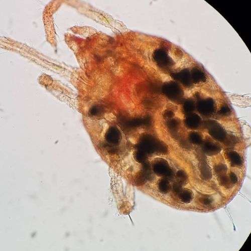
mite
Mite
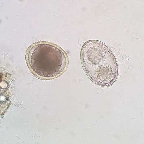
Toxocara canis (roundworm) on the left and Toxascaris leonina on the right.
Toxocara canis (roundworm) on the left and Toxascaris leonina on the right
Our Top 13 Microscopy Influencers To Follow
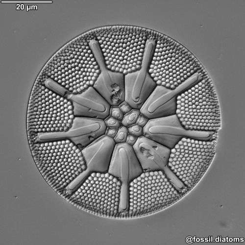
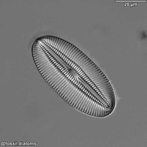
Diatom under the microscope with DIC illumination
Our Top 13 Microscopy Influencers To Follow
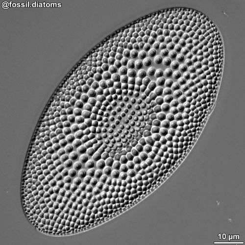
Diatom under the microscope with DIC illumination
Our Top 13 Microscopy Influencers To Follow
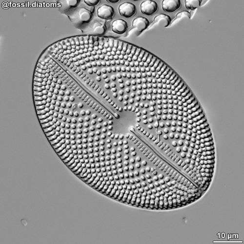
Diatom under the microscope with DIC illumination
Our Top 13 Microscopy Influencers To Follow
Diatom under the microscope with DIC illumination

JAKUB WITKOWSKI | @FOSSIL.DIATOMS

Subject matter: siliceous microfossils, mostly with diatoms, ranging in age from the Cretaceous to the Miocene (thus far). 

Microscope setup: I use a Zeiss setup with a 100x/1.4 oil DIC plan-apochromatic objective, and most of my photos are spliced using Helicon Focus.

“My name is Jakub Witkowski, I am an academic geologist based in Szczecin, north-western Poland. I work with siliceous microfossils, mostly with diatoms, ranging in age from the Cretaceous to the Miocene (thus far). I grew up with diatoms because they were the scientific occupation of my father – it became a family history of some kind.

I see my Instagram account as a way of science outreach. It includes diatom taxonomy, micropaleontology, evolution, all sorts of things related with biogenic silica. It is always a pleasure when people ask questions, and it came as the biggest surprise to me that diatoms draw so much attention from all kinds of artists.

Just recently, and in parallel to the Instagram Account, I established an online Fossil Diatom Atlas.”

Find Jakub on:

Instagram: @fossil.diatoms

Jakub’s Diatom Atlas: https://fossil-diatoms.com/atlas/

TRACY DEBENPORT | @UNDER.THE.SCOPE

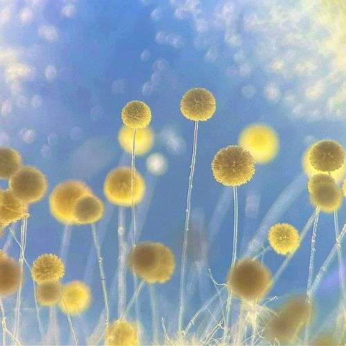
Subject matter: moulds and microbes

Microscope setup: Olympus CH2 compound microscope and Leica Stereozoom 4 microscope using a combination of an iPhone 11 and Nikon D3400 camera.

“My Instagram account features images that are meant to inspire curiosity and create positive associations with microbes. For example, many people think of pathogens and contaminants when they hear the word mold, but there are so many more beneficial molds then there are detrimental ones. Molds, and microbes in general, play vital roles in our ecosystems. They are amazing organisms and I try to bring attention to them by capturing and creating images that are beautiful, fun, and engaging.

I fell in love with microscopy and molds while working with Aspergillus flavus, a plant pathogen, at Cornell University. The first time I saw it under a microscope I was absolutely blown away. Its microscopic structures reminded me of flowers, pom-poms, and even peacock feathers. I had never considered that mold could look so beautiful, delicate, and familiar. I related to this organism in a whole new way. As I continued to explore the microscopic world of molds, I felt compelled to share images and information about these incredible organisms with friends and family, which is what lead me to start an Instagram account.

Today I’ve worked with both pathogens and beneficial microbes in a variety of areas such as agriculture, waste management and food.  I love that microbes are being studied more and more for their beneficial properties. A few examples being their potential to recycle materials, degrade toxic compounds, prevent disease in crops, produce new medicines, and serve as a sustainable and natural way to produce dyes for food and textiles.  There’s so much untapped potential, especially when it comes to fungi, and I hope that my images help to generate interest in, and appreciation of, these awesome organisms.”

Find Tracy on:

Instagram: @under.the.scope

Website: www.tracylovesfungi.com

Our Top 13 Microscopy Influencers To Follow
Aspergillus 3
Penicillium vulpinum
Veterinary cytology Francesco Cian
Francesco Cian
Veterinary cytology books
First and second edition of the Veterinary Cytology books by Dr. Kathleen Freeman, featuring Francesco Cian as co-author in the second edition

FRANCESCO CIAN | @VETERINARY_CYTOLOGY

Subject matter: veterinary cytology case studies

Microscope setup: Nikon Eclipse Ci with 4x, 10x, 20x, 40x, 50x oil and 100x oil objectives

“My interest for cytology started long time ago – in 2005 – when I was still a veterinary student. I spent a few months as an intern in the department of pathology. I developed fascination for looking at cells under the microscope and how this could be so important to identify the type of lesions (often tumours) that dogs/cats have and establish the most appropriate treatment.

When I graduated in 2007, cytology was a small side job as I was working in a busy small animal general practice. I then developed the desire to start a residency program in Veterinary Clinical Pathology, a broader discipline that includes cytology, haematology and clinical biochemistry. That’s the moment I felt I had the chance of my life. I was given by a friend a case-based cytology book from Dr. Kathleen Freeman that I found very inspiring (see pictures), and I started wondering if I could go and visit her for a few weeks. I found out she was working from home in a small village in Scotland in the middle of nowhere. I wrote her an email and very unexpectedly I got a reply very quickly saying that I could go at any time. With the money I got from a scholarship I won in Italy for the best thesis (on feline lymphoma), I booked my flights to Edinburgh and spent a month in Scotland. It was a fantastic experience that also opened the door for a residency in Veterinary Clinical Pathology at the University of Cambridge which I started in 2010. After I took the European Board (ECVCP) in 2014, I worked first at the Animal Health Trust and then at BattLab, a private diagnostic laboratory of the LABOKLIN group located in Coventry where I still am.  When I started my work at BattLab, Dr Kathleen Freeman asked me to work with her on the second edition of her cytology book, the one that inspired me originally as a young vet. I have to say that has been my biggest achievement so far and I felt I kind of closed a circle.

Over the years, I have also developed a major interest in teaching cytology to other vets. Cytology brought joy in my life, it opened a lot of opportunities and I felt the desire to share it with other enthusiastic people. I have travelled all over the world teaching cytology from China, Japan, Russia, Europe and USA. I have met – and hopefully inspired – thousands of veterinarians.

Nowadays online education is very popular, especially since the Covid pandemic. Even before that, I opened a veterinary cytology group, on Facebook first then on Instagram, that grew in popularity over the years and still is a place where vets from all over the world connect, share cases, cytology pictures and information about courses. We now have over 30000 members between the two platforms. I think it is a good example how technology can be beneficial and allow people to connect and share knowledge in a professional and healthy way.”

Find Francesco on:

Instagram: @veterinary_cytology

Facebook group: VeterinaryCytology

Website: veterinarycytology.org

Use this delivery method if you live in the Channel Islands. Delivery times 3-7 days typically.

No VAT will be charged on your purchase if the delivery address is to the Channel Islands.

All orders for stock items are dispatched within 2 working days using a 3-7 working day delivery service.

Please note, we regret that, because of the cost of delivery of items to your region, by selecting this delivery method, you agree that you will pay the difference in price (compared to UK rates) of any delivery charges for any replacement or repair within the warranty period. (If you do not wish to accept these terms please do not proceed with your order.)

All orders for stock items are dispatched within 2 working days using a 2-5 working day delivery service.

If the delivery charge is the same as the std mainland UK rate the condition below does not apply.

Please note if your delivery charge is higher than the std mainland UK rate that is because it is more expensive to deliver items weighing over 0.6Kg by courier to your region and the condition below therefore applies:

Please note, For any consignment weighing over 0.6Kg we regret that, because of the cost of delivery of items to your region, by selecting this delivery method, you agree that you will pay the difference in price (compared to std mainland UK rates) of any delivery charges for any replacement or repair within the warranty period. (If you do not wish to accept these terms please do not proceed with your order.)

Scottish Highlands means these postcodes as defined by our carriers: AB1-9/14/21-23/30-39/41-45/51-/56. IV1 – 56. KW1 – 14. KA27 – 28. PA20 – 39. PH11 – 40. FK19 – 21